Normal Floor Of Mouth Anatomy

Floor Of The Mouth The Intraoral And Extraoral Exam Continuing Education Course Dentalcare Com
Floor Of The Mouth

Oral Cavity Oncohema Key
Normal Oral Cavity Findings And Variants Of Normal Intechopen
Normal Anatomy Medtube Net

Parts Of The Mouth Staywell Api Web Examples

The floor of the mouth is examined using direct and indirect vision followed by bimanual palpation of the entire area.
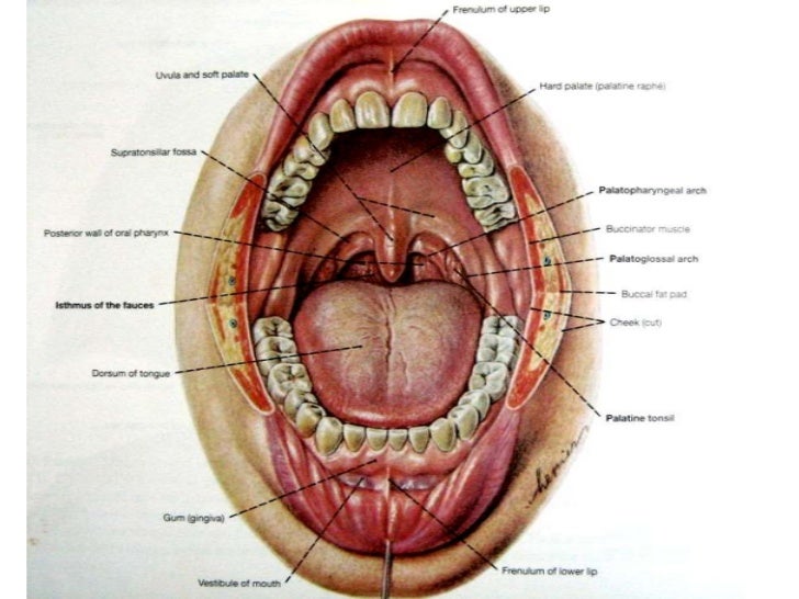
Normal floor of mouth anatomy. Geniohyoid muscles pull the larynx forward during swallowing. Cancer of the floor of mouth accounts for 28 35 percent of all mouth cancers. Food in the mouth is swallowed into the oropharynx and passed on to the esophagus and the rest of the gastrointestinal tract. The oral cavity leads to the oropharynx which begins at the anterior tonsillar pillars fig.
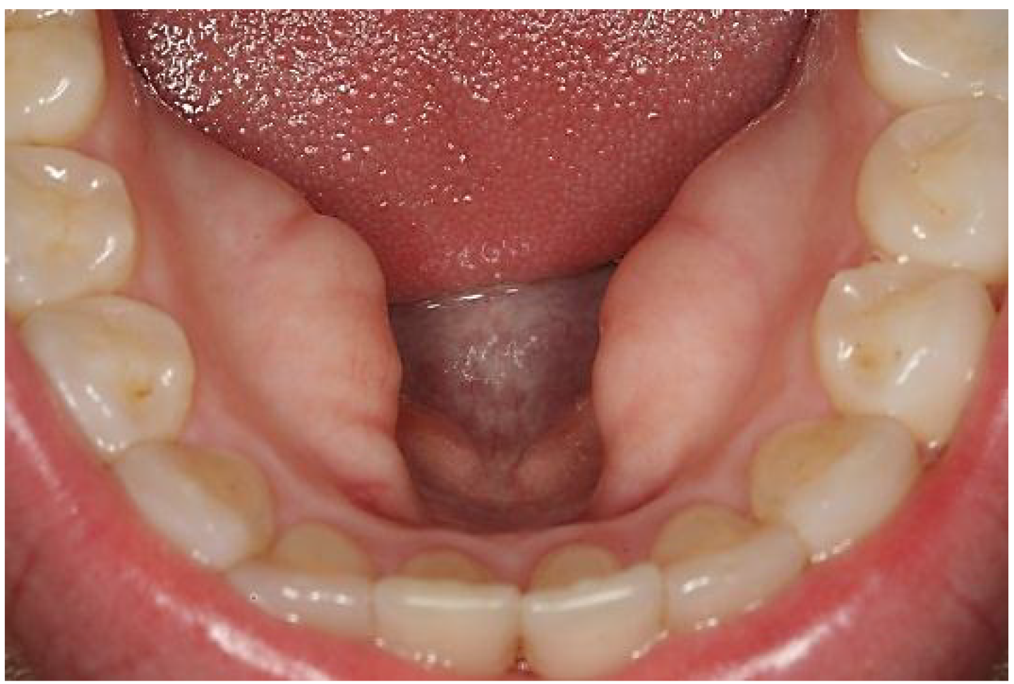
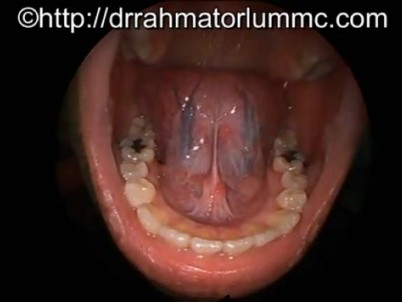
The nasopharynx starts behind the nose and lies above the oropharynx located at the very back of your mouth. It provides structural support to the floor of the mouth and pulls the larynx forward during swallowing. Lingual vein is superficially located and easily visible. Lymphoid aggregates are collections of normal or focally hyperplastic lymphoid tissue that may occur anywhere within the oral cavity but most commonly involve the regions of waldeyer s ring which includes the oropharynx lateral tongue soft palate and floor of mouth.
The floor of the oral cavity consists of several structures. The region of the throat behind the mouth is known as the oropharynx and forms the posterior wall of the mouth. The pharynx connects the nasal cavity and mouth to the esophagus and larynx in the neck. Anatomy of the floor of the mouth the floor of the mouth is a horizontally aligned u shaped space situated in the part of the oral cavity that lies beneath the tongue.
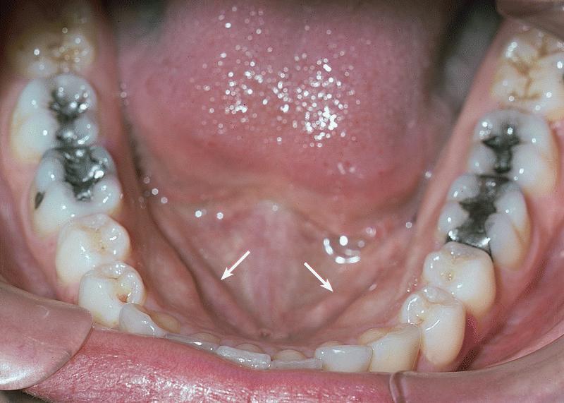
The patient should be asked to raise the tongue making direct visual examination of the tissues toward the midline of the floor of the mouth possible figure 44. For purposes of surgical planning the floor of the mouth is defined as the space between the mucosal surface and the mylohyoid muscle sling and comprising both structures 1. The nasopharynx the oropharynx and the laryngopharynx. The floor of mouth is a horseshoe shaped area under the tongue between the sides of the lower jawbone the mandible.
The pharynx or throat is a tube about five inches long composed of three parts. 190 2 190 5 and 196 1. These include the hard palate floor of the mouth buccal mucosa including the upper and lower gingivobuccal sulci retromolar trigone and anterior two thirds or oral portion of the tongue and lips figs. The submandbular gland is not visible but.
Visual examination of the floor of the mouth. Published on feb 13 2011 this video shows the normal anatomy of floor of mouth and the tongue. Men are diagnosed with floor of mouth cancer three to four times more often than women.
Pathology Outlines Anatomy

Human Mouth Wikiwand

Imaging The Floor Of The Mouth And The Sublingual Space Radiographics

Oral Cancer Screening Mhealth Org

Clinical Aspects Of The Sublingual Mass Note That The Normal Anatomy Download Scientific Diagram

Human Mouth Video Learning Wizscience Com Youtube

Floor Of The Mouth Flashcards Quizlet

Oral Cavity And Floor Of The Mouth Introduction Radiology Key

Sagittal T1 Mri Of Normal Oral Cavity Anatomy The Dashed Pink Line Download Scientific Diagram

Mouth Better Health Channel

Review Of Imaging Anatomy And Pathology Of The Floor Of The Mouth Semantic Scholar
The Oral Cavity
Http Pdf Posterng Netkey At Download Index Php Module Get Pdf By Id Poster Id 101807

